
Step by Step Visual Guide to Sketch Notes
Let Me Take You Visually Through Blood Circulation Sketch Notes
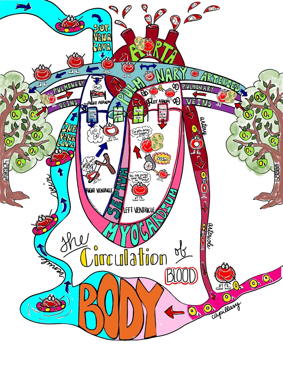
I know that using Sketch Notes is a different concept and can be a little scary if you’ve never done it before so let me take you step-by-step visually through an example. The blood circulation through the heart Sketch Notes are the most detailed ones I use with my students. My students get confused with which areas of the heart have oxygenated blood and which have deoxygenated. They also have trouble remembering that the pulmonary VEINS contain oxygenated blood from the lungs because this totally goes against what I have taught about arteries and veins. That’s why I made these Sketch Notes so detailed. They really help students visualize the process when they use their basic heart as a model. And, once you’ve seen the process I use for these complicated Sketch Notes with my anatomy students, simpler Sketch Notes will be a breeze for you to implement!Heart Structure & The Circulation of Blood
I drew these Sketch Notes the same way I taught my students to draw the heart easily (see original post with step by step instructions and video here). I think it really helps them to see things done the same way. Maybe it will help them remember it! (I hope so at least, it can’t hurt lol). These Sketch Notes are more detailed (intentionally, I promise 🙂 ) because it is always a topic that my students seem to struggle to remember. I’ve found that even my community college students trying to get into nursing school seem to get stuck on this sometimes. Since these are a little more “involved”, I thought it would be a good opportunity to share exactly how I am approaching these with my students in my own classroom.Here’s My Detailed Visual Guide for Using These Sketch Notes

I. Start Off with the Heart Structures
I will be using the version of the Sketch Notes with the vessel names already completed. My students have already worked a little with the basic structures of the heart so this is not new to them (they made “Anatomically Correct Valentines” see post here ). We will cover these structures in this order:- Myocardium- the heart muscle itself, it composes the walls of the heart
- Septum- the “wall” of muscle that divides the ventricles into a right & left half; this will be very important in our study of the Conduction System coming up next.
- Chambers– the heart is like a house in that it is divided into “rooms”, 2 on the top floor (atria or atrium is singular) and 2 on the bottom floor (ventricles). Since the chambers are like rooms, the valves are how you “exit” or leave the room– It helps students to think of it this way–> If you are staying within your house and simply traveling from room to room without going outside, you would use a door. If you would like to go outside but all the doors are locked, you would use a window. So, between “rooms” (chambers) are drawn as “doors” (the Bicuspid and Tricuspid Valves) and access to outside the heart (the major vessels) are drawn as “windows” (Pulmonary and Aortic Valves).
- Valves (Tricuspid, Bicuspid (Mitral), Pulmonary, Aortic)- I drew these as windows (going to the vessels) and doors (going between the atria and ventricles) so students would recognize that valves also allow entry into blood vessels and not just chambers (this is a common misconception with my high school students). We will also talk about what might happen in a person if the “door” or “window” valves didn’t close well (especially the Bicuspid (Mitral) Valve), or if there is a “door” between the atria (or a “hole” in the heart or atrial septal defect (ASD)). Both of these conditions are usually known by some of my students.
Major Vessels
- Major Vessels-
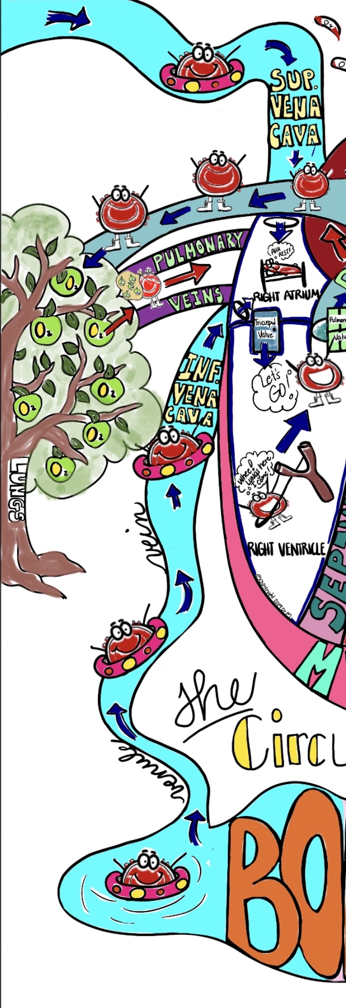
- Pulmonary Trunk/ Arteries- go to the lungs & pick up Oxygen (color these some version of blue because blood is deoxygenated here)
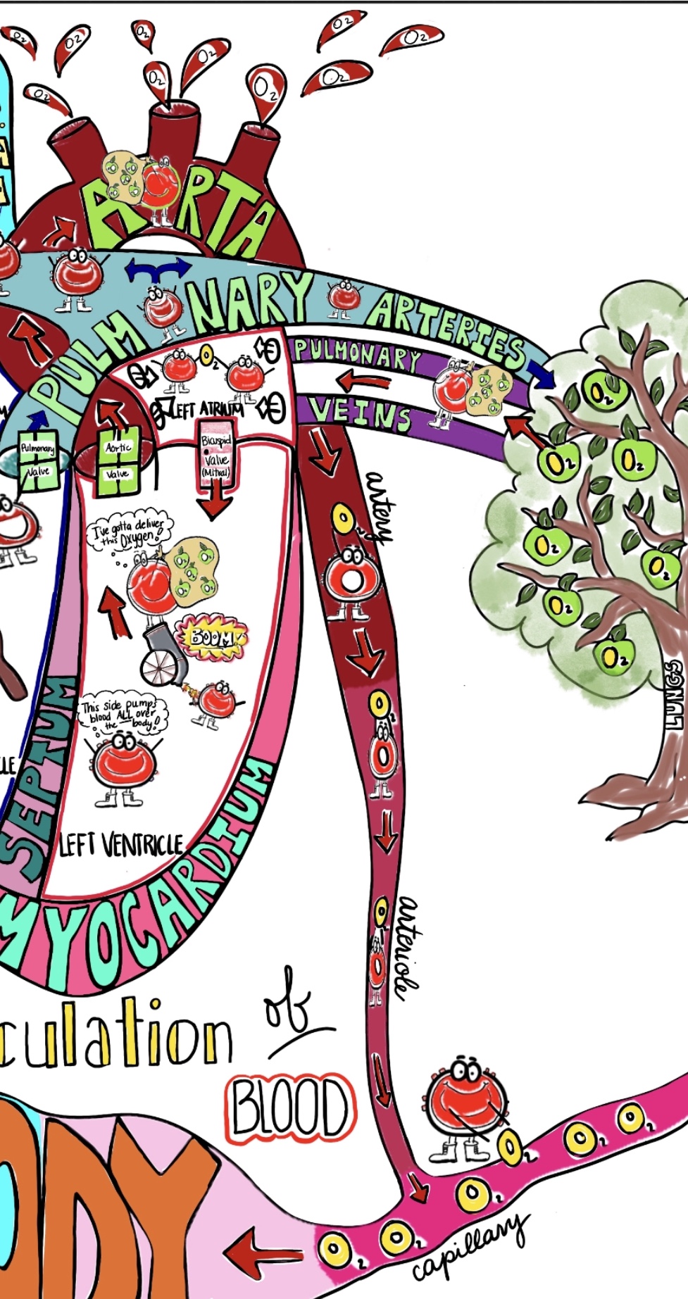
- Pulmonary Veins- return from the lungs with Oxygen & dump it into the Left Atrium (color these some version of red because blood is oxygenated here- I used purple because it is a mix of blue and red and these veins are exceptions to our rule)
- ** NOTE: The Pulmonary Arteries and Veins are EXCEPTIONS to the rule that arteries carry oxygenated blood away from the heart and veins carry deoxygenated blood towards the heart… This is often confusing to my students because they tend to remember artery=away (& if it’s leaving the heart, it’s carrying Oxygen to the tissues). There’s always an exception to every rule, and the Pulmonary Arteries & Veins are the exceptions to this one!
- Aorta- largest blood vessel in the body; takes oxygenated blood and pumps it ALL over the body so the blood in it is under tremendous pressure (so that’s why it’s being shot out of a cannon in the Sketch Notes). Blood that leaves the Aorta is carrying Oxygen (that’s what’s in his pack) and delivers it to the tissues of the body. (Color this a dark red color because blood is oxygenated here and we will vary the shades of red getting lighter as the vessels get smaller from artery to arteriole to capillary)
-
- Blood has to travel through increasingly narrow vessels– arteries (fairly large, maybe the diameter of a pencil; have thick muscular walls to contract and keep the blood moving!)–> arterioles (smaller, maybe the diameter of a spaghetti noodle; this is why the blood cell carrying O2 looks like he’s being squeezed!!)–> capillaries (TINY maybe the diameter of a single hair, red blood cells must line up single file to pass through them; this is where all the oxygen is delivered to the tissues in”capillary beds”; blood enters capillary beds WITH Oxygen (oxygenated) and leave capillary beds WITHOUT Oxygen (deoxygenated), this is the location that arteries transition into veins–> venules (small but larger than capillaries, about the same size as arterioles; these begin the journey of blood back to the heart)–> veins (largest, similar to the size arteries but walls are thin & don’t have to contract to move blood so it’s like a “lazy river” back to the heart. (On the picture, I forgot to vary the colors of blue for the venules& veins but you could do so if you want to show the difference in diameter)
- Superior Vena Cava (drains from the upper body) and the Inferior Vena Cava (drains from the lower body)- the 2 largest veins that channel blood into the Right Atrium (color these blue because blood in them is deoxygenated)
II. Then Discuss The Circulation of Blood

Tracing Path of Blood
Deoxygenated Blood
- I begin the path of blood in the Right Atrium. In the Right Atrium, the blood is not under high pressure, it is simply “chilling” (maybe not piled up in bed, but you get the idea lol 🙂 ) waiting on the chamber to fill so it can make the journey into the Right Ventricle. (Outline the Right Atrium & Ventricle in blue or shade in lightly because blood is deoxygenated in these chambers. Color any arrows on this side blue. Red blood cells in these chambers as well as the Pulmonary Trunk and Arteries should be a dark red color because blood that is deoxygenated is a crimson or dark red color)
- When the Right Atrium gets full, blood enters the Right Ventricle. If you think of the heart as a house, in order to get to another “room” (heart chamber), you would need to go through a door. The Tricuspid Valve is the “door” between the Right Atrium and Right Ventricle.
- Blood is not under exceptional pressure in the Right Ventricle, however, it does need a fairly strong muscular contraction to help it break a “window” (aka go through the Pulmonary Valve). I highly doubt a red blood cells slingshot themselves out of the heart lol, but it does represent that blood needs a substantial “push” (ventricular contraction) so it has enough momentum to break a “window” (go through the Pulmonary Valve), exit the Right Ventricle, and enter the Pulmonary Trunk and Arteries.
- Once blood passes through the Pulmonary Trunk and is diverted into the Right and Left Pulmonary Arteries, it begins it’s journey to the Lungs where it will “pick” up Oxygen molecules. I drew the Lungs as apple (oxygen) trees because it gives me an opportunity to introduce them to the bronchial tree and alveoli that we will study in the Respiratory System. We talk about how the bronchial tree is branched into smaller tubes until it ends in bunches of structures called alveoli. These alveoli look like the fruit (in this case apples containing Oxygen) at the end of branches on a tree. They are the site where diffusion happens and the oxygen we breathe in is handed over to red blood cells so they can deliver it to all of our body tissues. It is also the place where wastes (carbon dioxide) is transferred so it can be breathed out. I drew the “oxygen apples” so that students could visualize these molecules as they are “picked” up and carried to the tissues by red blood cells.
Oxygenated Blood
- Blood (carrying a pack full of “oxygen apples”) travels through the Pulmonary Veins from each lung until being emptied into the Left Atrium through 4 holes (2 on each side for each Pulmonary Vein). I remind students again of that the Pulmonary Arteries and Veins are the EXCEPTION to the “arteries carry oxygenated blood AWAY from the heart” rule because it is quite the opposite here! I drew red blood cells proudly holding up their Oxygen molecule in the Left Atrium so students would be reminded that this chamber contains oxygenated blood. (Outline the Left Atrium & Ventricle in red or shade in. Color the red blood cells on this side (& any with the pack) a bright red color since blood containing Oxygen is a bright red color)
- When the Left Atrium gets full of oxygenated blood, it enters the Left Ventricle through the “door” (Bicuspid or Mitral Valve). I remind students that problems with the Mitral Valve are a fairly common heart issue because sometimes the “door” doesn’t close completely or sometimes is warped and sticks. This causes the blood to leak into the “room” (chamber) when it shouldn’t. (ex. Mitral Valve Prolapse, Mitral Valve Stenosis)
- Once blood fills the Left Ventricle, the most vital part begins. The myocardium (muscle) MUST contract so forcefully that blood will have enough momentum to travel through vessels that range in size from the diameter of your finger (aorta), down to the diameter of a pencil (arteries), down to the diameter of a hair (arterioles), down to vessels so tiny blood cells line up single file to pass through them! … All while giving away their Oxygen to every tissue in our body! Talk about pressure!!! This is why I drew the red blood cell (with his pack of “oxygen apples”) being shot out of a cannon. The blood is under tremendous pressure as it leaves the Left Ventricle and shoots up the Aorta. To maximize it’s effectiveness, the Aorta has branches that divert the oxygenated blood to both the upper and lower portions of the body.
- Red blood cells deliver their Oxygen to capillaries in capillary beds through diffusion. These capillary beds are located all over the body so Oxygen distribution is most effective. I drew the blood cell tossing Oxygen molecules into a capillary “stream” which leads into a larger lake representing our whole body. After a wild ride so the red blood cells can deliver all of their Oxygen to the body, they can take it easy and simply float back (under low pressure) via veins to the heart. That’s why they are drawn floating in a swim ring on the opposite side of the “body”. Their trip returning to the heart would be more of a “lazy river” float compared to the high pressure trip they took via the arteries moving away from the heart.
- Large veins channel these easy-going red blood cells back to the heart from the upper part of the body (Superior Vena Cava) as well as the lower part of the body (Inferior Vena Cava). Blood enters the Right Atrium through 2 holes (1 at the top and 1 near the bottom of the outside) where it waits for the chamber to fill before starting the process all over again….. And all of this happens in under a minute! (and it could be longer or shorter depending upon which specific circulatory system it is traveling in 🙂 )
- Your heart beats 100,000 times a day, 35 million times a year. During an average lifetime, the human heart will beat more than three billion times. The adult heart pumps five quarts of blood each minute throughout the body – adding up to approximately 2,000 gallons of blood each day!
Extensions
My students will perform some extension activities with this unit. (They will have a choice between #2 and #3 but everyone will do #1)- Recall Race– They will try and see if they can recall the basic structures in the circulation of blood without looking. My students are pretty competitive, so we will race. As a final challenge, students will have to draw the quick heart (this one ) then label and draw in the circulation of blood.
- A Traveler’s Story- Using my Sketch Notes as an example, students will create their own story of the adventures of a red blood cell as it travels through the basic double circuit of circulation. They must include all structures and the final product can either be written as a creative story (with the structures highlighted so it’s easier to grade for me lol) or drawn as their own Sketch Note version.
- Stop Motion Video– Students will create a Stop Motion Video of the circulation of blood through the double circuit of circulation. They can use anything they want to simulate red blood cells, however, they must make a distinction between oxygenated and deoxygenated so their circulatory path is very clear. They must draw (& label!) the heart themselves (either on paper or on my desks or lab tables with chalk markers) to use for their video. Videos will be shared with me on Google classroom.
How to Learn More
I hope this is helpful for you (and your students)! To purchase these Sketch Notes to use with your class, visit https://www.teacherspayteachers.com/Product/Heart-Structure-Blood-Circulation-SketchNotesStudent-Notes-incl-FIB-Version-4401250 Please follow my TeachersPayTeachers store to receive updates when I upload additional Sketch Notes! https://www.teacherspayteachers.com/Store/Drm Also, don’t forget to visit my website www.biologysketchnotes.com to see some of my Biology Sketch Notes