
Let Me Show You How:
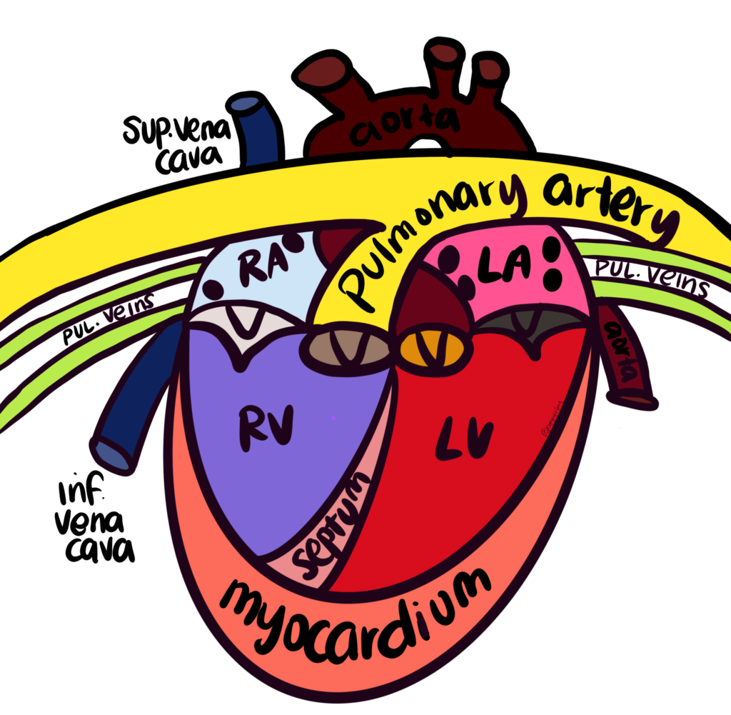
When we cover the structure of the human heart, I like to draw it for my students as we discuss it. This is an easy method that I learned one summer in an anatomy workshop. It is amazingly easy and my students rarely ever have trouble recalling the structure of the heart. You start by drawing 2 elementary school era “birds” connected by 2 circles. Like this:

Then, add in the Vs (V= Valves) 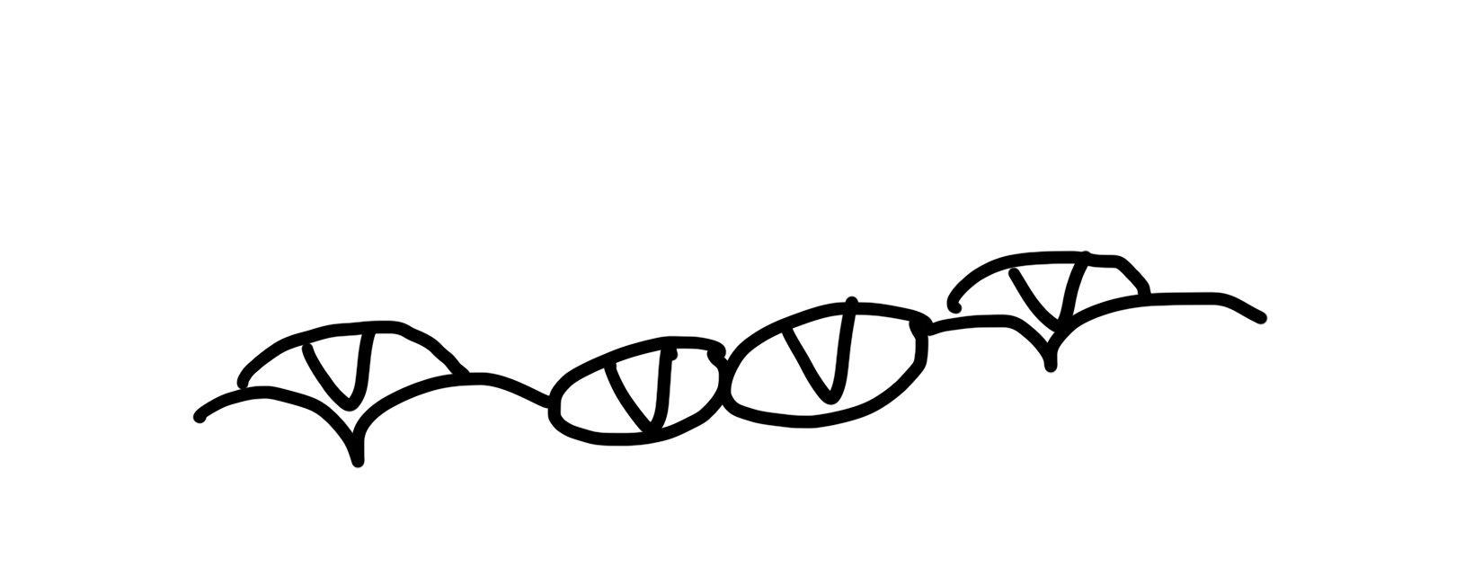
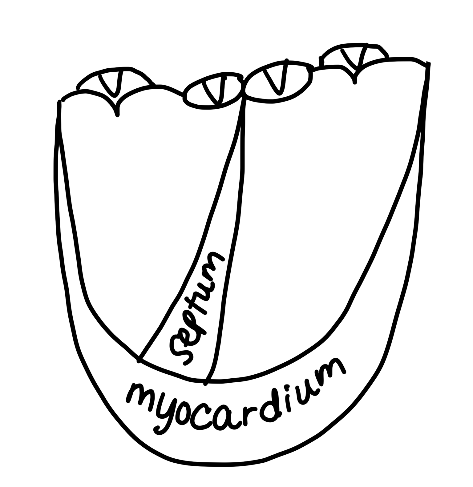
Connect the wings of the outer “birds” and create the myocardium (the muscular outer walls of the heart) and draw in the septum (division of the ventricles) between the 2 circles. 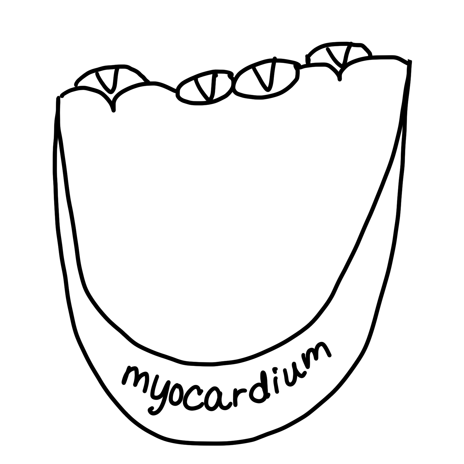
Off the first circle, create the pulmonary trunk and the pulmonary arteries. The pulmonary trunk is a major vessel of the human heart that originates from the right ventricle. It branches into the right and left pulmonary arteries, which lead to the lungs. Each of these vessels has elastic walls similar to those of the aorta, though somewhat thinner, and they are considered to be arteries even though the blood they carry is not oxygenated. The trunk itself is relatively short and wide. The function of these vessels is to transmit oxygen-depleted, carbon dioxide-rich blood from the right ventricle to the lungs. 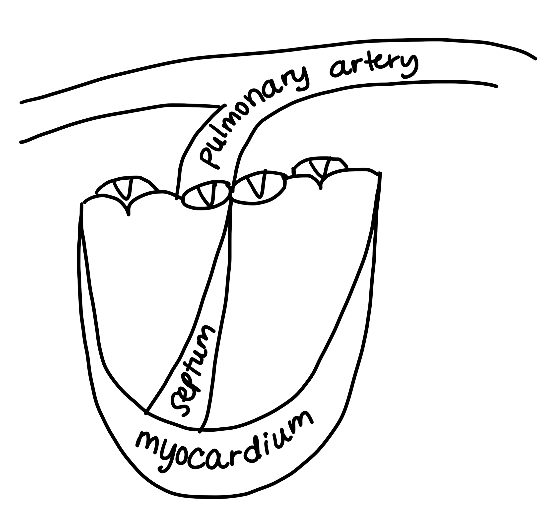
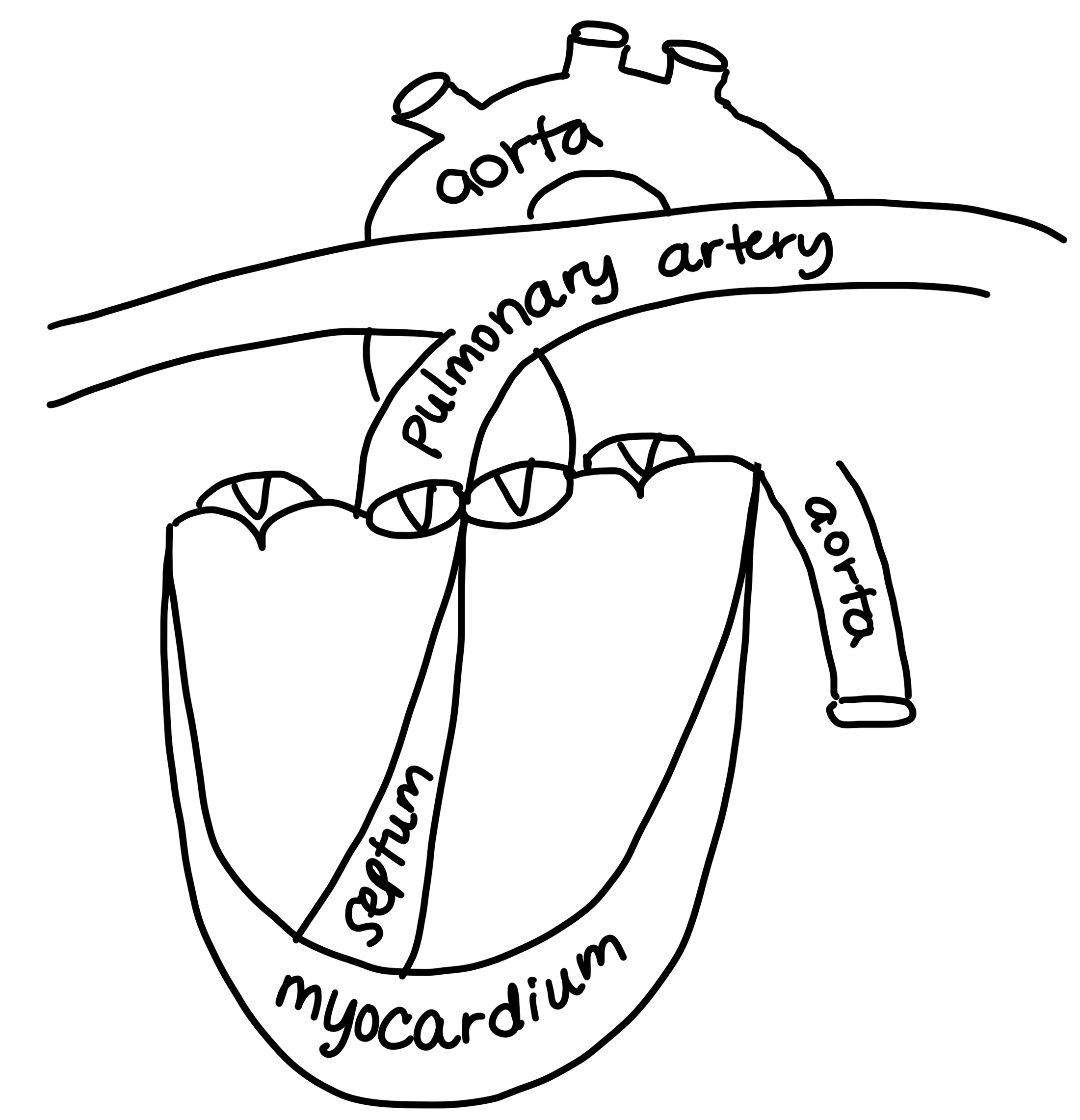
From the second circle, create the aorta which is situated behind the pulmonary trunk and arteries. The aorta is the largest artery in the body. The aorta begins at the top of the left ventricle, the heart’s muscular pumping chamber. The heart pumps blood from the left ventricle into the aorta through the aortic valve. Three leaflets on the aortic valve open and close with each heartbeat to allow one-way flow of blood.
The aorta is a tube about a foot long and just over an inch in diameter. The aorta is divided into four sections:
• The ascending aorta rises up from the heart and is about 2 inches long. The coronary arteries branch off the ascending aorta to supply the heart with blood.
• The aortic arch curves over the heart, giving rise to branches that bring blood to the head, neck, and arms.
• The descending thoracic aorta travels down through the chest. Its small branches supply blood to the ribs and some chest structures.
• The abdominal aorta begins at the diaphragm, splitting to become the paired iliac arteries in the lower abdomen. Most of the major organs receive blood from branches of the abdominal aorta.
Just under the pulmonary trunk and arteries, connect the lines to create the left atrium and right atrium. These are the top 2 chambers and receive blood returning to the heart from the body. Label the Right Atrium (R.A.), Left Atrium (L.A.), Right Ventricle (R.V.) and Left Ventricle (L.V). 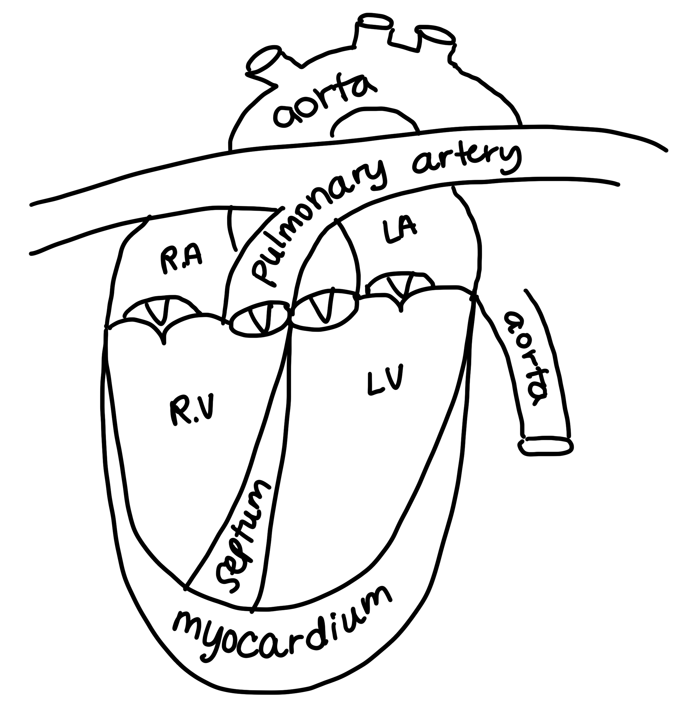
Connect the pulmonary veins to the Left Atrium on the right side and left side of the heart. Make sure to draw 4 dots to represent the places where the pulmonary veins return oxygenated blood from the lungs into the left atrium of the heart. 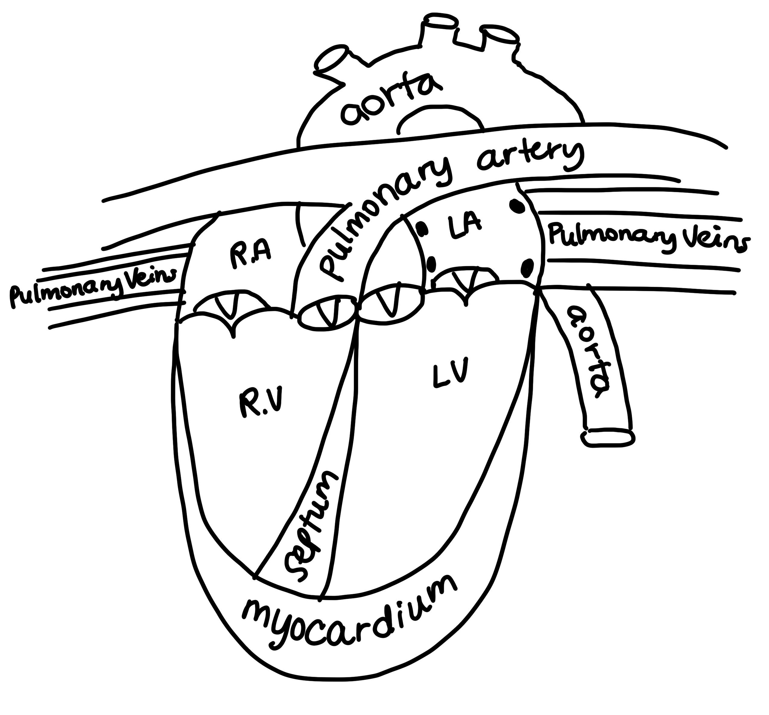
Draw the Superior Vena Cava above the Right Atrium and the Inferior Vena Cava next to the Right Ventricle. Make sure to draw 2 dots to represent the places where they return deoxygenated blood from the body into the Right Atrium. 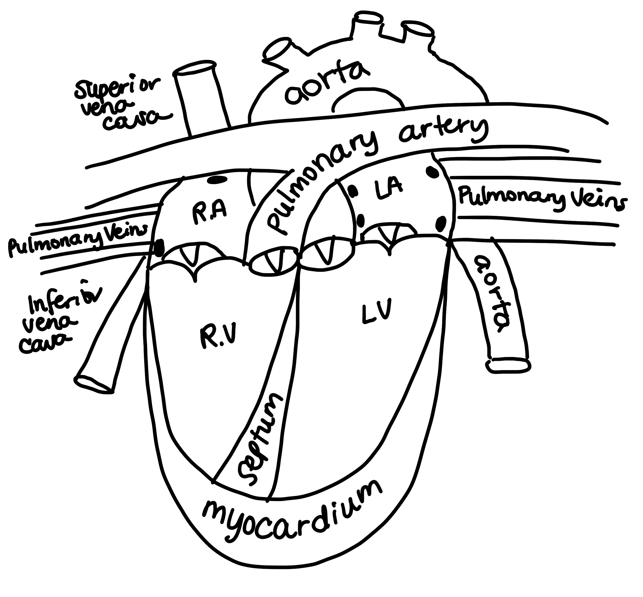
I draw this heart as we go over all the parts and have students draw along with me. Once we are finished, we set a timer and see who can draw it the fastest (correctness counts 🙂 ) Hope your students have fun with it!

3 Responses
This heart was so inspiring, this is what got me interested in going to med school.